


Contents
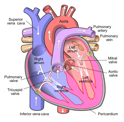
Inferior vena cava
The inferior vena cava is one of the main veins in the body.
Inferior vena cava: anatomy
Position. The inferior vena cava is mainly located in the abdomen.
Origin. The inferior vena cava arises at the level of the 5th lumbar vertebra. It corresponds to the union of the common iliac veins. (1) (2)
Path. The inferior vena cava runs up the front of the vertebral bodies and back of the aorta to the first lumbar vertebra. It then continues to rise, tilting to the right, and passes through the diaphragmatic orifice. (1) (2)
Termination. The inferior vena cava joins and ends at the level of the right atrium. (1) (2) At this level, a muscular fold is formed, called valve of the inferior vena cava or Eustachi valve.
Collateral branches. Numerous collateral branches open along the path of the inferior vena cava (1) (2):
- Lumbar veins. They form satellite veins in the lumbar arteries. Each lumbar vein ends at the back of the inferior vena cava.
- Renal veins. Forming two venous trunks, the renal veins open onto the lateral surfaces of the inferior vena cava at the level of the first lumbar vertebra.
- Right spermatic or ovarian vein. It goes up along the inferior vena cava before ending up below the opening of the renal veins.
- Right middle adrenal or capsular vein. It opens onto the rear face of the inferior cellar, between the opening of the renal veins and the passage through the diaphragmatic orifice.
- Hepatic veins. Usually two in number, these veins terminate in the inferior vena cava below the diaphragm.
- Lower diaphragmatic veins. They open onto the front face of the inferior vena cava, at the level of the diaphragmatic passage.
Venous drainage
The inferior vena cava leads venous blood to the heart, and more specifically to the right atrium (1) (2).
Pathologies and associated issues
Phlebitis. Also called venous thrombosis, this pathology corresponds to the formation of a blood clot, or thrombus, in the veins. These clots can move and move up to the inferior vena cava. This pathology can lead to various conditions such as venous insufficiency. The latter corresponds to a dysfunction of a venous network. When this occurs at the level of the inferior vena cava, the venous blood is then poorly drained and can impact the entire blood circulation (3).
Tumors. Benign or malignant, tumors can develop in the inferior vena cava. However, this cancerous development is uncommon (4) (5).
Trauma. Following a violent shock, the inferior vena cava can undergo trauma. This can be manifested by hypovolaemia, that is, a blood deficit. (4)
Treatments
Medical treatment. Depending on the pathology diagnosed, certain drugs may be prescribed such as anticoagulants or anti-aggregants.
Thrombolyse. Used during myocardial infarction, this treatment consists of breaking up the thrombi, or blood clots, with the help of drugs.
Surgical treatment. Depending on the pathology diagnosed, surgery may be necessary.
Chemotherapy, radiotherapy, hormone therapy, targeted therapy. Depending on the type and stage of the tumor, these treatments may be used to destroy cancer cells. (5)
Examination of the inferior vena cava
Physical examination. First, a clinical examination is performed to assess the symptoms perceived by the patient.
Medical imaging exam. In order to complete or confirm a diagnosis, a Doppler ultrasound, a CT scan, or an MRI can be performed.
History
Referred to as the Eustachi valve, the inferior vena cava valve is named after the famous 16th century Italian anatomist and physician, Bartolomeo Eustachi. (6)